change font size

How Often Should You See an Eye Doctor for a Comprehensive Eye Exam?

When was your last eye exam? If you’re unsure, you’re not alone. Many people delay scheduling an appointment until they notice changes in their vision. However, routine comprehensive eye exams are essential for maintaining optimal eye health, detecting problems early, and preserving clear vision throughout every stage of life. Understanding how often to visit an eye doctor can help you stay proactive about your health and prevent potential complications.


What Is a Comprehensive Eye Exam?

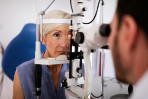
A comprehensive eye exam goes far beyond a basic vision screening. While screenings simply measure how well you see, a full examination evaluates both your visual acuity and the overall health of your eyes. During the appointment, an ophthalmologist assesses your prescription, eye coordination, and internal eye structures such as the retina and optic nerve.

Using advanced diagnostic technology, comprehensive exams can detect early signs of conditions like cataracts, glaucoma, macular degeneration, and diabetic retinopathy—often before noticeable symptoms develop. These thorough evaluations play a critical role in preventing vision loss and ensuring long-term eye health.

How Often Should You Schedule an Eye Exam?

The ideal frequency for comprehensive eye exams depends on your age, lifestyle, and medical history. While individual needs may vary, general guidelines include:

  • Children and Teenagers: Every 1–2 years to support proper vision development and academic success.
  • Adults Ages 18–39: Every 2 years if no vision problems or risk factors are present.
  • Adults Ages 40–64: Every 1–2 years as age-related changes and eye disease risks increase.
  • Adults Ages 65 and Older: Annually to monitor for conditions such as cataracts, glaucoma, and macular degeneration.
  • High-Risk Individuals: More frequent exams may be necessary for those with diabetes, high blood pressure, a family history of eye disease, or existing vision concerns.

An ophthalmologist can recommend a personalized schedule based on your specific needs.

Why Regular Eye Exams Matter

Routine eye exams are about more than just updating your glasses or contact lens prescription. Many serious eye conditions develop gradually and without symptoms, making early detection essential. Comprehensive exams allow for timely treatment, reducing the risk of permanent vision loss.

In addition to safeguarding your eyesight, eye exams can reveal signs of broader health concerns such as diabetes, hypertension, and high cholesterol. Because the eyes provide a unique view of blood vessels and nerves, they often serve as an early indicator of systemic conditions.

Advanced Eye Care You Can Trust

Patients seeking expert care can rely on Sierra Nevada Eye Center. Specializing in ocular surgery and the treatment of ophthalmic disorders, the practice utilizes state-of-the-art technology to deliver exceptional patient care. With facilities in Reno, Renown, and Carson City, their experienced team provides comprehensive eye exams and advanced treatments tailored to each individual.

Protect Your Vision for the Future

So, how often should you get a comprehensive eye exam? The answer depends on your age, health, and risk factors—but regular visits are essential for preserving lifelong vision. By prioritizing routine eye care, you can detect issues early, maintain clear sight, and protect your overall well-being.

If it’s been a while since your last exam, now is the perfect time to schedule one, or give us a call, and take a proactive step toward healthier vision.

If you’ve been diagnosed with cataracts, you may be considering cataract surgery to restore clearer vision and improve your quality of life. Cataract surgery is one of the most common and successful procedures performed today, and with the right preparation, you can help ensure a smooth and comfortable recovery.

At Sierra Nevada Eye Care, patients in Carson City and Reno benefit from advanced surgical techniques and a team of specialists dedicated to personalized care. Whether this is your first cataract surgery or you’re exploring your options for the first time, understanding how to prepare can make a significant difference in your experience.


Schedule a Comprehensive Preoperative Exam

Preparation for cataract surgery begins with a comprehensive eye exam. Your eye doctor will measure your eye’s structure, evaluate your overall eye health, and discuss your vision goals. This evaluation helps determine the best type of intraocular lens (IOL) for your needs, whether it’s a standard lens or a premium option that can reduce dependence on glasses.

Understand Your Intraocular Lens Options

One of the most important decisions in cataract surgery is choosing the right IOL. There are several types available, including monofocal, multifocal, and toric lenses, each designed to address specific vision needs.

Your eye surgeon will explain the benefits and limitations of each lens and recommend the best option for your lifestyle. For example, patients who enjoy reading, driving, or outdoor activities may benefit from lenses that reduce the need for glasses after surgery. Understanding your options beforehand ensures you have realistic expectations and are prepared for the recovery process.

Follow Pre-Surgery Instructions Carefully

Before your surgery, your eye doctor will provide detailed instructions to prepare your eyes and body for the procedure. Common recommendations include:

  • Avoiding certain medications: Blood-thinning medications may need to be paused temporarily. Always follow your doctor’s guidance.
  • Using prescribed eye drops: Some patients receive antibiotic or anti-inflammatory drops to reduce the risk of infection.
  • Fasting if required: Depending on the anesthesia used, you may need to refrain from eating or drinking for a specific period before surgery.

Following these instructions closely helps minimize complications and ensures your surgery goes smoothly. Patients from both Reno and Carson City have found that careful adherence to preoperative instructions leads to faster, more comfortable recoveries.

Understand What to Expect During Recovery

Recovery from cataract surgery is usually quick and straightforward, but it’s important to have realistic expectations. Most patients notice improved vision within 24 to 48 hours, though full stabilization may take several weeks.

During recovery, you may experience:

  • Mild irritation or scratchiness
  • Blurry or fluctuating vision
  • Sensitivity to light

Your eye doctor will provide a schedule for follow-up visits to monitor healing and ensure no complications arise. Using prescribed eye drops consistently and avoiding rubbing or putting pressure on your eyes are crucial for a smooth recovery.

In the first days after surgery, it’s important to avoid activities that could strain your eyes or increase the risk of injury. This includes:

  • Heavy lifting or strenuous exercise
  • Swimming or hot tubs
  • Eye rubbing or applying pressure to the eyes
  • Extended screen time in some cases (follow your doctor’s guidance)

By minimizing strain, you can help your eyes heal properly and reduce the risk of complications.

Prepare Your Home for Recovery

Setting up a comfortable recovery environment at home can make a significant difference in your post-surgery experience. Consider the following tips:

  • Clean and declutter: Clear walkways to reduce the risk of bumps or falls.
  • Stock up on necessities: Have medications, eye drops, snacks, and water within easy reach.
  • Set up a relaxing space: Comfortable seating, good lighting for reading, and easy access to entertainment can help pass the time during the first few days of recovery.

Maintain Follow-Up Appointments

Follow-up visits with your eye surgeon are essential to ensure your eyes are healing correctly and your vision is improving as expected. During these visits, your doctor will check for signs of infection, monitor intraocular pressure, and verify that your vision aligns with your goals.

Stay Positive and Patient

Finally, maintaining a positive attitude can make the recovery process easier. Cataract surgery is highly effective, but your eyes need time to adjust. Most patients are thrilled with the results and notice a significant improvement in clarity and color perception within a few days. By preparing properly, following your surgeon’s instructions, and giving your eyes time to heal, you’ll set yourself up for the best possible recovery and enjoy the benefits of clearer vision for years to come.


Take Our Free Vision Assessment

Are cataracts affecting your daily life? Our quick self-assessment can help you understand when it might be time for surgery.

Common signs it’s time for cataract surgery:

  • Difficulty reading, even with strong lighting
  • Struggling with night driving or glare from headlights
  • Colors appearing dull or yellowed
  • Frequent changes in prescription glasses
  • Double vision or halos around lights

Schedule a Consultation at Sierra Nevada Eye Care

If you’re considering cataract surgery, scheduling a consultation with a trusted eye care team is the first step. Sierra Nevada Eye Care offers expert care for patients in Carson City and Reno, with advanced technology and personalized guidance for every stage of the cataract surgery process.

By understanding how to prepare for cataract surgery, you can reduce anxiety, minimize complications, and enjoy a smooth recovery. Clearer vision may be closer than you think—take the first step today and schedule your consultation to discuss your options with our experienced team.

Schedule Your Comprehensive Cataract Evaluation Today

Do words on the menu or your phone look blurry? Has it become increasingly difficult to see? 

You might have a cataract. Cataracts are typically not noticeable at first, but you may start to experience symptoms like blurred, cloudy vision and poor night vision as the cataract grows. 

Some of the symptoms of a cataract can be the same as those of presbyopia, so they can be hard to diagnose on your own. Keep reading to learn more about cataracts, including seven signs you may be experiencing cataracts instead of presbyopia!

What Is the Difference Between Presbyopia and Cataracts?

You can have presbyopia and cataracts at the same time. However, they’re completely different eye conditions.

What is Presbyopia?

Presbyopia happens when your eye lens becomes less flexible as you grow older. So, how does that affect your vision?

Your lens allows you to focus. It changes shape to provide clear, up-close vision.

But with age, the lens loses its flexibility. When your lens is rigid, it can’t change its shape the same way. 

Consequently, it becomes difficult to focus on close-up objects. Most people start to notice the effects of presbyopia at around age forty. 

Common symptoms of presbyopia include:

  • Squinting 
  • Difficulty reading small print
  • Trouble focusing on near objects
  • Eye fatigue from doing close activities
  • Needing brighter lighting for up-close work
  • Headaches or eyestrain after reading or performing close work
  • Holding reading materials at arms-length to make words clearer  

What is a Cataract?

Like presbyopia, a cataract also affects your lens. But cataracts occur when proteins in your eye break down and clump on your lens, causing it to become cloudy. 

Most cataracts grow gradually and won’t disturb your vision in the early stages. You may not even realize you have a cataract until it’s more advanced. 

Over time, cataracts lead to poor vision and may eventually cause blindness. But any vision lost to cataracts can be regained through cataract surgery. 

Cataracts may form in one or both eyes. When they’re in both eyes, they often develop at different rates.

What Are the Most Common Signs of Cataracts?

You may have a cataract if you are experiencing any of the following symptoms:

1. Blurry Vision

Does your eyesight become blurry when reading a text or looking at an object? It could be a sign of a cataract. 

Blurred, filmy, or cloudy vision is one of the most common symptoms of cataracts. As a cataract becomes bigger and more opaque, it blocks light entering your eye, causing blurry vision. 

It may feel like you’re looking at the world through a foggy window when you have cataracts. With time, your vision might become blurry, making watching TV, driving, and performing other routine activities increasingly difficult.

2. Sensitivity to Light

You may experience light sensitivity when you have a cataract. A clear lens lets light pass easily through it. 

The lens then works together with your cornea to focus light properly onto the back of your eye or retina. But when you have a cataract, the cloudy lens scatters light and prevents it from focusing correctly on the retina. 

This can cause light sensitivity. As a result, bright lights and the sun might cause eye pain or make you squint or close your eyes.

3. Frequent Prescription Changes

Rapidly changing vision is a tell-tale sign of cataracts. As your cataract worsens, you’ll find that you need a stronger prescription often. 

A different strength of glasses or contact lenses can improve your eyesight, but it’s only a temporary fix. Eventually, you’ll need cataract surgery to restore clear vision.

4. Needing Brighter Lighting to See Clearly

If you require more light to read a book or brighten your phone screen to see images more clearly, you may have cataracts. When a cataract darkens your lens, brighter lighting can help you see clearly for a while. 

Once this stops working, your eye doctor may recommend cataract surgery.

5. Halos and Glare

You may have cataracts if you’ve noticed increased glare from lights and halos around light sources. The presence of a cataract can lead to diffraction of light entering your eye, resulting in halos and glare.

6. Poor Night Vision

Patients with cataracts often complain of worsening nighttime vision. Cataracts tend to darken or dim your vision as they progress.

These changes may not be apparent during the day when there’s sufficient natural light to compensate for your declining vision. However, dimming vision becomes more evident at night. 

Combined, dim vision, halos, and glare can make nighttime driving dangerous to you and other road users. Seeing halos around streetlights and street signs while driving can be bothersome and distracting. Glare from oncoming headlights may become blinding.

If you’re having trouble with your night vision, it’s important to stop driving at night and inform your eye doctor. 

7. Colors Appearing Faded

Cataracts can make colors appear washed out, dull, or faded. Cataracts will turn brown or yellow as they mature. 

This discoloration is passed onto your vision, causing objects to take on a yellowish or brownish hue. As a result, it may become hard to tell certain colors apart, like blue, purple, and green, or black, navy blue, and brown. 

If you often disagree with family and friends about the color of a shirt or dress, it’s time to see your eye doctor.

Get to the Bottom of Your Symptoms

The only way to know for sure if you have cataracts or presbyopia is to see the experienced doctors at Sierra Nevada Eye Center. If you have a cataract, our doctors will monitor it and let you know when it’s the right time for surgery.

Do you have signs of cataracts? Schedule your cataract screening at Sierra Nevada Eye Center in Reno, NV, today for an accurate diagnosis and proper management.

Cataracts are the leading cause of vision loss in the U.S. Over time, cataracts can grow to significantly impact your vision and even cause blindness if left untreated.

Fortunately, vision can be easily restored through a simple procedure.

With cataract surgery, you can get rid of cataracts for good and restore clear vision.

Cataract surgery is a quick, outpatient procedure that can even allow you to experience greater visual freedom. Keep reading to learn more about cataracts, including how cataracts affect vision!

What are Cataracts?

Cataracts develop when proteins inside the natural lens of your eye begin to break down and clump together. These changes cause your lens to become cloudy and make it increasingly difficult for light to pass through. 

If the light is unable to pass through the lens to reach the retina, vision will be blurry. Many people with cataracts say it’s like looking through a foggy window. 

What Are the Symptoms of Cataracts?

Cataracts typically develop so gradually that you might not notice any vision changes. But as they become more advanced, you may experience the following symptoms:

  • Blurry, foggy, or cloudy vision 
  • Sensitivity to light 
  • Poor night vision
  • Halos and glare
  • Colors looking faded
  • Double vision in one eye 
  • Frequent prescription changes
  • Needing brighter light for close-up activities

Why Cataract Surgery is Necessary?

In the early stages of cataracts, new prescription glasses or contact lenses, brighter lighting, and magnifying lenses can help you see better. However, cataracts will worsen with time. 

These solutions stop working when your cataract grows bigger and covers a more significant part of your lens. Cataract surgery is the only effective treatment for cataracts. 

Your eye doctor will recommend cataract surgery once your symptoms begin to interfere with your daily life. It is important not to wait until your vision is significantly impacted to have cataract surgery.

This is because hyper-mature cataracts that lead to blindness are harder to remove and increase the risk of complications during surgery. So, it’s best to have cataract surgery before cataracts cause total vision loss.

Usually, your eye doctor will recommend cataract surgery if you can’t watch TV, use your phone, drive safely, cook, read, and perform other routine activities. 

What Happens During Cataract Surgery?

During your cataract procedure, eye drops will be administered to widen your pupil. Numbing drops will also be applied to completely numb your eye.

You’ll remain awake during the entire procedure and see general movement and light. However, your eye doctor will ensure that you are comfortable.

From there, your cataract surgeon will create a tiny incision in your cornea. The opening will allow your cataract surgeon to access your cloudy lens. 

Next, your cataract surgeon will break the cataract into tiny pieces and remove them. Once all the pieces are removed, your cataract surgeon will implant an artificial lens or intraocular lens, also known as an IOL, to take the place of your natural lens. 

The new IOL is what restores clear vision after cataract surgery. Depending on the IOL you choose before your cataract procedure, you could end up with even greater visual freedom than before. 

Finally, the tiny incision made is left to heal on its own without stitches. 

If you have cataracts in both eyes, your cataract surgeon will likely wait until the treated eye heals first before removing the cataract in the other eye. 

What Recovery Looks Like After Cataract Surgery

Your cataract surgeon will provide aftercare instructions before you leave. It’s very important to follow all of them closely for a safe and smooth recovery.

Since cataract surgery is an outpatient procedure, you will be able to go back home on the same day of your procedure. You will need to arrange for someone to drive you to and from the surgery center in advance.

Your eye doctor will prescribe anti-inflammatory and antibiotic eye drops to reduce inflammation and prevent infection. You’ll also have scheduled post-op appointments. 

Make sure you attend all your follow-up visits. These appointments allow your eye doctor to ensure your eye is healing properly.

Recovery following cataract surgery is usually quick. A majority of patients resume most of their routine activities within one to three days after surgery.

How Does Cataract Surgery Improve Vision?

Here are some of the ways cataract surgery can improve your eyesight:

Clear Vision

Removing cataracts clears up your vision. When your cloudy lens is replaced with a new one, images and objects become sharper and crisper than before.

Some IOLs can provide clear vision at all distances: near, far, and everything in the middle. Others can correct refractive errors like presbyopia and astigmatism. 

Depending on the IOL you select, you may be able to reduce your dependency on visual aids after the procedure. Best of all, cataracts can never come back once they are removed. 

So you can look forward to enjoying clear vision for years to come.

Brighter Colors

One of your most unforgettable moments after cataract surgery will be realizing what colors you’ve been missing. Colors tend to lack luster and look washed out when you have cataracts. 

This is because you view the world through a yellowish or brownish lens instead of a clear one when you have cataracts. As a result, everything around you takes on a brownish or yellowish hue. 

After cataract surgery with a clear IOL, you’ll experience a beautiful range of colors. Suddenly, colors will appear brighter and more vivid.

Reclaim Your Vision from Cataracts

If you’re unable to perform basic tasks like cleaning or cooking or even do the things you love, like golfing, reading, or hiking, because of cataracts, you should consider cataract surgery at Sierra Nevada Eye Center. Cataract surgery will restore your vision and significantly improve your quality of life.

Has your vision changed enough to interfere with your everyday life? Schedule your cataract screening at Sierra Nevada Eye Center in Reno, NV, today to find out if it’s time for cataract surgery!

Glaucoma is one of the leading causes of irreversible blindness worldwide. It’s called the silent thief of sight because it doesn’t usually have any symptoms in the early stages until significant vision loss has occurred. 

Fortunately, you can preserve your remaining vision and live a full life when glaucoma is detected and treated early. Keep reading to learn more about glaucoma and laser treatments for this progressive eye condition!

What is Glaucoma?

Glaucoma is a group of eye conditions that often cause a buildup of fluid in your eyes. The accumulation of fluid increases your eye pressure or intraocular pressure, also known as IOP. 

Over time, the high eye pressure damages your optic nerve. The optic nerve sends messages from your retina to your brain to create the images you see. 

Damage to the optic nerve causes permanent vision loss or blindness.

What is Open-Angle Glaucoma?

Inside the front section of your eye is a clear fluid called the aqueous humor. Your eye is constantly creating this fluid. At the same time, an equal amount of the fluid drains out through the trabecular meshwork in the drainage angle. 

The trabecular meshwork is the spongy tissue near the edge of your cornea. It allows the aqueous humor to flow out of your eye.

Your eye pressure rises when fluid can’t drain properly or there’s an overproduction of the aqueous humor. This is what happens in open-angle glaucoma.

Open-angle is the most common form of glaucoma. It tends to progress very slowly. So, there are usually no symptoms during the early stages. 

By the time you notice any changes in your vision, it’s often late in the disease, and extensive damage has already occurred. In the late stages of open-angle glaucoma, you may experience the following symptoms:

  • Patchy vision loss in your peripheral or side vision
  • Trouble seeing things in your central vision

What is Angle Closure Glaucoma?

Angle-closure or closed-angle glaucoma is a rare but serious type of glaucoma that needs immediate medical attention. It develops when your cornea and iris move toward each other, closing the angle between them.

The closed angle completely blocks off the fluid’s access to your eye’s drainage system, which comprises the trabecular meshwork and uveoscleral drains, preventing the aqueous humor from flowing out. When the flow is suddenly blocked, fluid builds up rapidly and causes a dangerous rise in eye pressure. 

Closed-angle glaucoma is an emergency that can cause irreversible blindness in only a matter of hours. It requires prompt treatment to reduce damage to your optic nerve and preserve your remaining vision. 

If you experience any of these symptoms, seek medical attention right away:

  • Severe eye pain
  • Blurred vision
  • Severe headache
  • Eye redness
  • Nausea or vomiting
  • Halos around lights

What is Normal-Tension Glaucoma?

Sometimes, the optic nerve becomes damaged even when eye pressure remains within normal levels. This is known as normal tension or low-tension glaucoma.

Eye doctors still don’t know exactly why some people develop normal tension glaucoma. However, a lack of blood flow to the optic nerve or a very delicate optic nerve that could be easily damaged by normal eye pressure may be factors in this form of glaucoma.

Are Laser Treatments Available for Glaucoma?

The damage already caused by glaucoma can’t be reversed. However, there are effective treatment options that can slow or stop further vision loss, especially if glaucoma is caught early.

The goal of any glaucoma treatment, including laser treatment, is to lower intraocular pressure and slow the progression of glaucoma. Here are some of the most common laser treatments for glaucoma:

Laser Trabeculoplasty

Your eye doctor may suggest laser trabeculoplasty if you have primary open-angle glaucoma. The laser targets your trabecular meshwork, opening it up. 

In turn, this increases fluid outflow and brings your eye pressure down. This simple yet highly effective procedure takes around five minutes to complete. 

On average, laser trabeculoplasty lowers eye pressure by around twenty to twenty-five percent. Your eye doctor will need to assess the effectiveness of the treatment during your follow-up visits.

Laser Iridotomy

The fluid known as aqueous humor normally exits the eye through the trabecular meshwork after passing through the angle. However, if the angle is narrow, the fluid encounters difficulty in its passage, leading to its accumulation and the subsequent buildup of pressure within the eye.

Laser iridotomy treats narrow-angle or angle-closure glaucoma by creating a tiny hole in your iris using a laser. The new opening provides an alternative channel for fluid to flow out, which relieves the pressure inside your eye.

Endocyclophotocoagulation (ECP)

Endocycyclophotocoagulation is typically performed with cataract surgery to decrease pressure inside your eye. It works by delivering laser energy to the ciliary body. 

The laser energy alters the cells in the ciliary body so that they produce less fluid. You’ll have to wait about four to six weeks to know if the treatment worked.

Transscleral Cyclophotocoagulation (CPC)

Transscleral cyclophotocoagulation is another type of laser treatment used to reduce your intraocular pressure. The laser energy is applied to the ciliary body so that it produces less aqueous humor. 

During the procedure, the laser probe physically touches your sclera, which is the white of the eye. Then, it transmits laser energy to the ciliary body. 

Decreasing the amount of fluid the ciliary body produces helps lower intraocular pressure. The quick outpatient procedure lasts approximately three minutes. 

Like other glaucoma laser treatments, your eye pressure won’t go down immediately, so you’ll be examined by your eye doctor about a week after treatment.

Don’t Let Glaucoma Steal Your Sight

In most cases, comprehensive eye exams with your eye doctor at Sierra Nevada Eye Center is the only way to detect glaucoma early. Early detection and treatment will increase your chances of slowing the progression of this sigh-threatening condition and preserving your vision.

Is it time for you to be screened for glaucoma? Schedule an appointment at Sierra Nevada Eye Center in Reno, NV, today!

Cataracts are a common eye condition that can cause a variety of visual symptoms, including blurry vision. They develop when the eye’s natural lens becomes cloudy or hardened. 

Typically, cataracts develop gradually and don’t cause noticeable changes in your eyesight at first. But as time passes, you may feel like you’re looking through a foggy window.

Keep reading to learn more about cataracts, including whether or not they are age-related!

What is a Cataract?

Cataracts occur when proteins in your eye break down and clump together inside your lens. Over time, a cataract continues to grow, causing more visual symptoms.

Typically, your eye’s natural lens is clear. It has the very important job of focusing the light that enters your eye onto the retina, enabling you to see crisp, sharp images. 

When the lens develops a cataract, it blocks light entering your eye and makes it gradually difficult to see clearly.

What Are the Signs of Cataracts?

Since cataracts tend to grow slowly, you might not notice any changes in your vision initially. But over time, you may experience the following symptoms:

  • Blurred, cloudy, or foggy vision 
  • Poor night vision
  • Halos and glare
  • Sensitivity to light 
  • Colors appearing faded or dull
  • Frequent prescription changes
  • Double vision in one eye
  • Need for brighter light for reading and other up-close activities

What Causes Cataracts?

Age is the most significant risk factor for cataracts. Experts believe that your body’s ability to fight the effects of oxidation on your lens decreases as you grow older. 

In response to oxidative stress, the proteins in your eyes start to deteriorate, break down, and accumulate on your lens, contributing to cataract formation. Age-related cataracts may begin to develop at around age forty. 

However, most people experience cataract symptoms by the time they reach sixty. Certain factors can cause you to develop cataracts earlier in life, such as:

Genetics

It is thought that genetics may play a role in the early development of cataracts. A family history of cataracts puts you at a higher risk of getting them earlier than usual.

Eye Trauma

An eye injury can lead to the clouding of your lens. This is called a traumatic cataract. 

A traumatic cataract is usually due to a penetrating or blunt force injury to your eye, which damages the lens fibers. Some of the common causes of a traumatic cataract include:

  • Head injury
  • Chemical burns
  • Infrared lights
  • Electric shock

Traumatic cataracts develop soon after your eye injury or years later.

Diabetes

Uncontrolled blood sugar in patients with diabetes is linked to the early formation of cataracts. Having high blood sugar for long periods can damage the blood vessels in your eyes. 

In turn, the damaged blood vessels can raise your chances of developing cataracts. Also, if you have high glucose levels over an extended period, enzymes in your eye lens may convert the glucose to sorbitol. 

Sorbitol can make your lens swell, contributing to blurry vision.

Prolonged Use of Corticosteroids

Corticosteroids are medications used to reduce inflammation. However, long-term use of corticosteroids prescribed for blood disorders, severe allergies, and other health conditions can speed up the development of cataracts. 

Lifestyle

Certain lifestyle choices can accelerate the formation of cataracts. They include:

  • Smoking
  • Excessive drinking
  • Too much exposure to the sun’s UV rays 

What Are the Treatment Options for Cataracts?

When cataracts are first developing, prescription glasses or contact lenses and stronger lighting can help you see clearly when you have cataracts. But eventually, your eye doctor will recommend surgery if poor vision from cataracts interferes with everyday activities like driving, reading, cooking, or watching TV. 

Cataract surgery is the only way to get rid of cataracts for good and restore clear vision. During cataract surgery, your cataract surgeon will remove the cloudy lens and replace it with a new artificial lens called an intraocular lens (IOL). 

The artificial lens implant is designed to remain permanently in your eye. There are different types of IOLs, including those that can provide clear vision at all distances. 

Your ophthalmologist will discuss the various IOL options available before your procedure and help you choose one that meets your vision needs and lifestyle.

Can You Prevent Cataracts?

There’s no research that shows how to prevent cataracts. However, eye doctors suggest the following strategies to reduce your risk of developing cataracts early:

Quit Smoking

If you smoke, you can ask your doctor to help you stop.

Maintain Healthy Glucose Levels

For people with diabetes, it’s important to keep sugar levels in check to lower the risk of getting cataracts.

Wear Sunglasses

Wear high-quality sunglasses that block 99 percent to 100 percent of UVA and UVB rays. Also, invest in wide-brimmed hats to ensure your eyes are protected from all sides when you’re outdoors.

Eat Right

Add a variety of leafy greens and colorful fruits into your diet, including avocados, bell pepper, citrus fruits, broccoli, carrots, green peas, red grapes, kiwifruit, sweet potatoes, kale, and spinach. All these have essential nutrients and antioxidants that support your eye health.

Reduce Alcohol Intake

You don’t have to give up drinking altogether. Limiting your alcoholic beverages to one drink per day can decrease your risk of cataracts.

Avoid Corticosteroids

Although corticosteroids are very beneficial, care should be taken when using them. If you’re taking corticosteroids, discuss the potential risks with your healthcare provider.

Ask if there other treatment options instead or avoid using corticosteroids for prolonged periods.

Get Regular Eye Exams

Frequent eye examinations at Sierra Nevada Eye Center are essential in reducing the risk of cataract-related vision loss and maintaining your eye health. The experienced eye doctors at Sierra can detect a cataract early, closely monitor it, and recommend surgery before it significantly affects your vision.

Are you experiencing symptoms of cataracts? Schedule an appointment at Sierra Nevada Eye Center in Carson City, NV, today!

Glaucoma is a group of eye diseases that gradually damage the optic nerve at the back of the eye. The optic nerve is responsible for carrying visual information from the eye to the brain so you can see. 

Glaucoma typically has no symptoms in its early stages. So without timely detection and prompt treatment, damage to the optic nerve can lead to permanent vision loss.

Keep reading to learn more about glaucoma, its causes, and the available treatment options!

What Causes Glaucoma?

Often, glaucoma is caused by an increase in your eye pressure, also known as intraocular pressure or IOP, which can cause damage to the optic nerve. Your eye constantly produces a clear fluid known as the aqueous humor.

The aqueous humor nourishes your eye and maintains your eye shape. It then leaves your eye through the drainage angle.

If there’s any blockage in your drainage angle, the rate at which your eye produces the aqueous humor becomes greater than the rate at which it can drain it, leading to high intraocular pressure. A buildup of pressure in your eyes damages the optic nerve causing permanent vision changes.

At the same time, some people can have high intraocular pressure without glaucoma, a condition called ocular hypertension. Others can also develop glaucoma even with normal eye pressure. 

Certain factors can increase your risk of getting glaucoma. They include:

  • Being age forty and older
  • Previous eye injury or surgery
  • Family history of glaucoma
  • Prolonged use of corticosteroids
  • Extreme nearsightedness or farsightedness
  • Some health conditions, such as diabetes, high blood pressure, sickle cell anemia, and heart disease

What Are the Types of Glaucoma?

There are many types of glaucoma. Some examples are:

Primary Open-Angle Glaucoma

Open-angle glaucoma is the most common form. It happens your eye’s drainage canals become blocked or clogged, and fluid is unable to leave the eye at a consistent and healthy rate.

As a result, fluid drains very slowly through the trabecular meshwork, gradually increasing your eye pressure. The trabecular meshwork comprises small channels where the aqueous humor flows out of your eye.

There are typically not many obvious symptoms of open-angle glaucoma in the beginning stages. For this reason, it’s important to get regular eye exams so the condition can be detected early.

Angle-Closure Glaucoma

Also called closed-angle glaucoma, this form of glaucoma occurs when the angle of your eye, which is an area the fluid has to flow through before exiting the eye, is closed off. Angle-closure glaucoma is less common and can happen suddenly. 

The drainage angle is where your iris and sclera, the white of your eye, meet. Sometimes, the iris can block the drainage angle and prevent fluid from draining properly, increasing your eye pressure. 

If you have symptoms of angle-closure glaucoma, seek treatment immediately. Left untreated, angle-closure glaucoma can cause permanent vision loss.

Some common symptoms of angle-closure glaucoma are redness, severe headache or brow ache, blurred vision, eye pain, and nausea or vomiting.

Normal-Tension Glaucoma

In normal-tension glaucoma, the optic nerve becomes damaged when your eye pressure is within the normal range. Many times, this means that your eye pressure is still too high for your individual eyes. 

What Are the Treatment Options for Glaucoma?

In many cases, the treatment method will depend on the type of glaucoma and its severity. While glaucoma doesn’t have a cure, treatment can slow or stop its progression. 

The treatment options for glaucoma include:

Medicated Eye Drops

The first line of treatment for glaucoma is usually prescription eye drops. Eye drops work by either increasing the outflow of fluid from your eye or reducing the amount of fluid produced by your eye. 

Some eye drops are able to do both. Decreasing the amount of fluid in your eye slows optic nerve damage and limits vision loss.

Laser Treatment

Your eye doctor may recommend laser treatment if eye drops don’t adequately lower your eye pressure. There are various types of laser procedures.

An iridotomy uses a laser to create tiny holes in your iris. These holes let the fluid drain more easily from your eye.

Another laser procedure used to treat glaucoma is selective laser trabeculoplasty, or SLT. During SLT, your eye doctor uses a laser to open clogged drainage canals in the trabecular meshwork. 

Removing the obstructions using a laser allows the aqueous humor to drain easily once again.

Glaucoma Surgery

Glaucoma surgery is necessary in advanced cases or when other treatment methods do not work to lower your eye pressure. Your eye doctor at Sierra Nevada Eye Center can also recommend surgery if eye drops cause serious side effects.

One type of glaucoma surgery is trabeculectomy. Trabeculectomy is a procedure done to create a new pathway in the sclera. The new path lets fluid leave your eye, which lowers your intraocular pressure and prevents vision loss from worsening.

At Sierra Nevada Eye Center, we are proud to offer minimally invasive glaucoma surgeries or MIGS. MIGSs are procedures that use microscopic equipment and tiny incisions to decrease your eye pressure in order to minimize or prevent optic nerve damage.

There are different types of MIGS procedures, including Kahook Dual Blade, iStent Inject, OMNI GATT, and Hydrus microstent. If you have mild to moderate glaucoma, MIGS can be a good option. 

The goal of MIGS procedures is to lower intraocular pressure in the following ways:

  • Reducing the production of the aqueous humor
  • Improving the eye’s natural drainage system
  • Redirecting excess fluid outside of your eye.

Protect Your Sight from Glaucoma

Glaucoma can sneak in without any warning signs and rob you of your sight. Luckily it’s treatable if caught early through regular eye exams at Sierra Nevada Eye Center.

Do you want to learn more about glaucoma or determine if you might be at risk of developing the condition? Schedule an appointment at Sierra Nevada Eye Center in Reno, NV, today!

Do your eyes often feel dry? Does it feel as though there’s always something in your eye? 

These could be signs of dry eyes. Dry eyes can affect your vision, make it hard to perform simple, daily activities, and get in the way of enjoying everyday life. 

Luckily there are various at-home treatments that can help with your dry eyes. Keep reading to learn more about dry eyes, home remedies for dry eyes, when to see an eye care professional, and the various treatment options available for dry eyes!

What is Dry Eye?

Dry eyes occur when your eyes produce poor-quality tears or don’t make enough tears. When you have dry eyes, you may experience the following symptoms:

  • Itchiness
  • Eye fatigue
  • Light sensitivity
  • Blurry or double vision 
  • Difficulty driving at night
  • Burning or stinging sensation
  • Trouble wearing contact lenses
  • Stringy mucus around or in your eyes
  • Feeling like there’s something in your eyes

Are There Home Remedies for Dry Eyes?

Although home remedies will not always be enough to resolve your dry eye symptoms, there are some things you may be able to do to help reduce them. Here are some tips for managing dry eyes at home:

Use Artificial Tears

Artificial tears lubricate your eyes, providing temporary relief from dry eyes. You may have to apply these eye drops multiple times throughout the day since the effects only last temporarily.

When buying over-the-counter artificial tears, look for preservative-free eye drops. That’s because eye drops with preservatives are more likely to irritate your eyes and worsen your symptoms.

Take Frequent Screen Breaks

Spending too much time in front of your computer, laptop, tablet, smartphone, or TV can lead to digital eye strain. You can combat eye strain and dry eyes from excessive screen time by following the 20-20-20 rule.

According to the rule, take a break from digital screens after every 20 minutes. Look at something at least 20 feet away for 20 seconds. Also, remember to blink often during your breaks. Blinking intentionally is a great way to lubricate your eyes and ease the symptoms of dry eyes.

Add Omega-3 Fatty Acids to Your Diet

Consider incorporating omega-3 fatty acids into your diet to aid with your dry eyes. Omega-3 fatty acids keep your eyes healthy and lubricated.

Some rich sources of omega-3 fatty acids include soybeans, walnuts, chia seeds, flaxseeds, and cold-water fish like salmon and tuna. Eating foods packed with omega-3 fatty acids can improve your tear quality and reduce your dry eye symptoms.

Stay Hydrated

Your tears are ninety-eight percent water. So ensure you remain hydrated throughout the day to help your eyes with tear production and alleviate your dry eye symptoms. 

Aim to drink the recommended amount of six to eight glasses of water per day at intervals.

Invest in a Humidifier

Dry air can cause dry eyes. Using a humidifier in your home or office adds much-needed moisture, which can aid with the symptoms caused by a dry environment.

Wear Wraparound Sunglasses

Wraparound sunglasses protect your eyes from the sun and wind, which can exacerbate your dry eye symptoms. Ensure you get wraparound sunglasses that fit snugly without causing any discomfort.

When to See an Eye Doctor for Your Dry Eyes

At-home treatments usually help with dry eye symptoms. But if your symptoms become worse or persist even with at-home remedies, it’s time to see an eye care professional.

An experienced eye doctor can determine what’s causing your dry eye and provide the best treatment to relieve your dry eyes.

Your dry eye treatment options may include:

Prescription Eye Drops

There are many types of eye drops used for treating dry eyes. Xiidra and Restasis are two of the most commonly prescribed eye drops. 

Both medications offer significant relief if you have moderate to severe dry eyes. It can take months before you really notice a difference when using Restasis. 

On the other hand, Xiidra starts working within two weeks, but it takes about three months to experience its full effect.

Punctal Plugs

If your tears leave your eyes too quickly, causing dry eyes, your doctor may recommend punctal plugs. Punctal plugs are small, biocompatible devices implanted in your tear ducts or puncta to block drainage during a procedure called punctal occlusion.

Consequently, tears remain on the surface of your eyes longer and provide more lubrication, which significantly improves your dry eye symptoms. Punctal plugs can be temporary or permanent. 

Temporary plugs dissolve on their own after several weeks. If temporary punctal plugs work well, your doctor may suggest permanent plugs.

Permanent plugs are made of acrylic or silicone and don’t dissolve. However, they can be removed if necessary.

Prokera 

Prokera is a non-invasive device that is similar to a contact lens. It has anti-scarring and anti-inflammatory properties that help your eyes heal.

Prokera is made from two transparent, flexible rings that sandwich a tiny piece of amniotic membrane tissue. During the same-day procedure, your ophthalmologist starts by applying anesthetic drops. The numbing drops reduce any potential discomfort.

Your ophthalmologist will then insert Prokera over your cornea, similar to how a contact lens is placed. The amniotic membrane comes from a natural placenta and can help heal your cornea from dry eyes.

Find Lasting Relief from Dry Eyes

If at-home remedies don’t alleviate your dry eye symptoms, the experienced eye doctors at Sierra Nevada Eye Center can help. The doctors offer comprehensive dry eye treatment, including prescription drops, punctal occlusion, and Prokera for long-term relief from dry eyes.

Are you struggling with the frustrating symptoms of dry eyes? Schedule an appointment at Sierra Nevada Eye Center in Carson City, NV, today to find a lasting solution!

Are cataracts making it hard to complete simple, routine tasks? Has poor vision from cataracts made your favorite pastimes less enjoyable? 

If so, your eye doctor will likely recommend cataract surgery. There are two options for removing your cataracts: traditional or laser cataract surgery. 

Traditional cataract surgery uses handheld blades and is a highly effective procedure. On the other hand, cataract surgery with laser technology is bladeless, offers unmatched precision, and delivers predictable outcomes.

Keep reading to learn more about laser cataract surgery, including eight things you may not know!

How Does Laser Cataract Surgery Work?

Cataracts occur when proteins in your eyes break down and clump together on your lens, making your vision cloudy, blurred, and discolored. Often, cataracts progress gradually and eventually cause blindness without treatment. 

Cataract surgery is the only way to restore vision affected by cataracts and get rid of them permanently. During cataract surgery, your cataract surgeon will remove your cloudy natural lens. 

Then, they will implant an artificial intraocular lens (IOL) in its place to restore clear vision. Cataract surgery using laser technology can ensure your procedure is more successful. 

LenSx femtosecond laser and ORA wavefront technology are among the latest techniques used in cataract surgery. Wavefront eye mapping allows your surgeon to take real-time, precise measurements and make necessary adjustments at any point in surgery.

In turn, this increases your chances of achieving the best possible vision post-surgery without contacts or glasses. If you’re struggling with poor vision from cataracts, you can regain your sight and live life to the fullest with cataract surgery. 

Here are eight reasons why you should consider getting laser cataract surgery:

1. Greater Precision

Laser technology creates a detailed, 3-D map of your eye surface. From the information gathered, like the size, location, and depth of the cataract, your surgeon will know exactly where to make the incision.

Surgical incisions with a computer-aided laser are up to 10 times more precise than incisions done by hand.

2. Safer Capsulotomy

Capsulotomy is a vital step in cataract surgery. It involves opening the front part of the delicate, thin membrane or capsule that holds your natural, cloudy lens. 

It’s crucial that the portion left inside your eye remains intact. That way, it can hold the new artificial lens in the correct position for clear vision.

Unlike traditional cataract surgery, where the capsule is opened manually, laser cataract surgery utilizes a computer-guided laser to open the capsule. The laser is able to create a more centered and circular opening in your capsule. 

Even the most experienced surgeons can’t match the precision of a computer-controlled laser.

3. Uses Less Energy to Break Up Cataracts

During laser cataract surgery, the surgeon uses a laser to soften the cloudy cataract lens. With the cataract softened, less ultrasound energy is required to break and remove it. 

Less energy reduces the risk of complications like corneal swelling and clouding or retinal detachment post-surgery.

4. Less Damage to Surrounding Tissue

Breaking up your cataract with a laser uses much less ultrasound energy. As a result, the surrounding tissues don’t suffer as much collateral damage as they would with traditional cataract surgery. 

This helps ensure your cornea remains clear and the lens capsule isn’t weakened.

5. Better Vision 

Even a slight misalignment of your new intraocular lens can decrease visual quality. The perfect opening made in your capsule by a laser optimizes your surgeon’s ability to accurately place your IOL and attain the best visual outcome. 

Thus, you’re more likely not to need contact lenses or glasses after your laser cataract procedure.

6. Can Correct Astigmatism

Astigmatism is a common refractive error. It occurs when your cornea has an egg shape like a football rather than being round like a baseball. 

The irregular curvature of the cornea causes blurry or distorted vision at all distances. Your surgeon can treat your astigmatism by reshaping the irregular cornea with a laser. 

Laser technology enables your surgeon to perform astigmatism correction more accurately and reliably, leading to better vision outcomes. By removing cataracts and fixing astigmatism simultaneously, many patients are able to see better than they have in years.

7. Minimally Invasive

Bladeless laser cataract surgery doesn’t use stitches. After removing your cataract and implanting an intraocular lens, the surgeon leaves the tiny incisions to heal on their own. 

A no-blade, no-stitch procedure is less-invasive and more comfortable for patients.

8. Easier Recovery

Since laser cataract surgery doesn’t use blades, your eye experiences less trauma. Laser cataract surgery also requires less energy to break up your cataract, reducing post-surgery corneal swelling.

Additionally, the clean, accurate incisions made using a laser reduce the risk of infection. All these things combined allow for a smoother recovery and faster vision restoration.

Cataract Surgery with the ORA System

Sierra Nevada Eye Center is proud to offer the ORA system and LenSx femtosecond laser, the latest in cataract surgery. The ORA system and femtosecond laser allow the experienced cataract surgeons at Sierra Nevada Eye Center provide truly customized procedures with excellent outcomes.

Are you interested in laser cataract surgery? Schedule your cataract screening at Sierra Nevada Eye Center in Carson City, NV, today to find out if it’s time for cataract surgery and whether laser cataract surgery is right for you.

Glaucoma is a group of eye diseases that damage your optic nerve. The optic nerve transmits images from the retina to your brain, allowing you to see. 

Damage to the optic nerve is irreversible and causes vision loss. If you’ve been diagnosed with glaucoma, prompt treatment is critical to protect your remaining vision and prevent permanent blindness.

Keep reading to learn more about glaucoma and how treatment helps with the condition!

How Common Is Glaucoma?

Glaucoma is the leading cause of irreversible blindness in the world. According to the Centers for Disease Control and Prevention (CDC), approximately three million Americans have glaucoma.

There are different types of glaucoma. The most common form typically progresses very slowly and usually has no early symptoms. 

That’s why many people with glaucoma don’t know they have the condition. It’s crucial to have regular eye exams to ensure glaucoma is caught and treated early before significant vision loss occurs.

What Are Some Types of Glaucoma?

Two types of glaucoma are angle-closure and open-angle glaucoma.

Open Angle Glaucoma

Open-angle is the most common type of glaucoma. It affects over ninety percent of glaucoma patients, advances very slowly, and often has no early physical symptoms.

Open-angle glaucoma occurs when a blockage in your eye’s drainage system causes fluid buildup. An accumulation of fluid increases your eye pressure and damages the optic nerve.

Angle-Closure Glaucoma

Acute angle-closure glaucoma leads to an abrupt increase in your eye pressure or intraocular pressure, also known as IOP. It develops quite quickly and is a medical emergency. 

Acute angle-closure glaucoma can cause rapid vision loss and blindness without treatment. Symptoms of angle-closure glaucoma include:

  • Severe eye pain
  • Halos around lights
  • Severe headache
  • Blurred vision
  • Eye redness
  • Nausea and vomiting  

Are There Risk Factors for Glaucoma?

Anyone can get glaucoma. However, some people are more at risk than others. The risk factors for glaucoma are:

  • Age over fifty-five
  • Thinner corneas 
  • Family history of glaucoma
  • Previous eye surgery
  • Serious eye injury in the past
  • Hispanic, African American, or Asian heritage
  • Long-term use of corticosteroid medications
  • High intraocular pressure
  • Extreme farsightedness or nearsightedness
  • Certain medical conditions like high blood pressure, diabetes, heart disease, sickle cell anemia, or migraines
  • Lifestyle choices like smoking, excessive alcohol use, or too much exposure to the sun

Eye doctors recommend getting a comprehensive eye exam every year if you have any of the above risk factors. Regular eye examinations are key to preventing unnecessary vision loss.

What Types of Glaucoma Treatments Are Available?

Glaucoma still doesn’t have a cure. But with careful monitoring and treatment, it can be effectively controlled and vision loss prevented. 

The goal of glaucoma treatment is to lower your eye pressure. Reducing your intraocular pressure slows the progression of glaucoma and helps preserve your eyesight. 

Glaucoma treatment is usually required for life.

Eye Drops

Eye drops are usually the first line of glaucoma treatment. They reduce your eye pressure by either improving the flow of fluid through the drainage angle or decreasing the amount of aqueous fluid produced by the eye.

Eye drops are applied several times daily, sometimes in combination with oral drugs. If your glaucoma worsens or your medication doesn’t lower your eye pressure to safe levels, your doctor may need to change your treatment over time.

Laser Surgery

In addition to eye drops, your glaucoma specialist may suggest laser surgery to treat your condition. One laser procedure that is common in those with narrow-angle or angle-closure glaucoma is an LPI or laser peripheral iridotomy.

During the LPI procedure, our eye doctor applies a local anesthetic to numb your eye. Then, they will use a laser to create a tiny hole in the iris, which is the colored part of your eye.

The new opening allows the aqueous fluid to drain out your eye more easily and increases the angle size. Increased outflow brings your eye pressure down and slows or prevents optic nerve damage.

For open-angle glaucoma, selective laser trabeculoplasty, or SLT, is used to decrease the production of fluid in your eye. It can also be used to increase the outflow of fluid from your eye.

Following your laser procedure, you may experience mild blurry vision and irritation. But you should be able to resume your normal routine the same day.

The type of laser procedure your doctor recommends will depend on the kind of glaucoma you have and its severity.

Glaucoma Surgery

You may need glaucoma surgery if:

  • You can’t tolerate eye drops
  • Other treatment options don’t work
  • You have acute angle-closure glaucoma or advanced glaucoma, where a blockage causes a rapid increase in your eye pressure

Surgery is urgently required to intervene and protect your remaining vision. There are different types of glaucoma surgeries, they include:

  • Trabeculectomy
  • ExPress mini-tube shunt
  • Ahmed and Baerveldt tube shunts 
  • Canaloplasty
  • Minimally invasive glaucoma surgeries (MIGS) – iStent Inject, OMNI, Kahook Dual-Blade, Hydrus Microstent, and GATT.

Prevent Vision Loss from Glaucoma

The top-rated glaucoma specialists at Sierra Nevada Eye Center recommend frequent eye exams. That’s the only way to detect glaucoma early and ensure you maintain a lifetime of the best vision possible.

Are you experiencing symptoms of glaucoma or want to have your eyes screened for the condition? Schedule an appointment at Sierra Nevada Eye Center in Reno, NV, today!

CONTACT US